Pesquisa
US Quiz of the Month – July 2020
CASE REPORT
A 51 year-old woman with left iliac fossa discomfort underwent a colonoscopy. Her past medical history only included a breast fibroadenoma diagnosed 4 years earlier. Physical examination and laboratory testing were unremarkable. Colonoscopy revealed a 50 mm polypoid lesion, with normal overlying mucosa, occluding the lumen at 20 cm from the anal verge, compatible with a subepithelial lesion (Fig. 1).

Figure 1. Colonoscopy: a polypoid lesion, with normal overlying mucosa, occluding the lumen.
An endoscopic ultrasound (EUS) was performed, showing a 49×32 mm hypoechoic, round, well-demarcated lesion arising from the muscularis mucosa (Fig. 2).
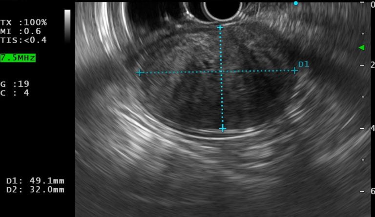
Figure 2. EUS: a hypoechoic, round, well-demarcated lesion.
EUS-elastography revealed a predominantly hard lesion with dispersed heterogenic soft areas (Fig. 3).
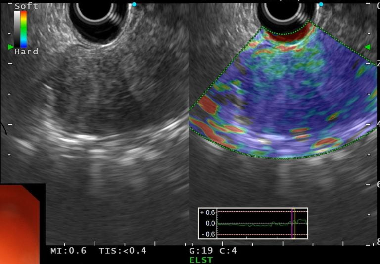
Figure 3. EUS elastography: a predominantly hard (blue) lesion with dispersed heterogenic soft (green and red) areas.
A EUS-guided fine-needle aspiration was performed using a 22-gauge needle.
WHAT IS THE MOST LIKELY DIAGNOSIS?
DISCUSSION
Hystological examination showed spindle cells arranged in bundles, with dense distribution of nucleus, forming palisades in dense fibrillar stroma. Immunohistochemical stains revealed that the tumor cells were positive for S-100 and negative for CD117, CD34 and a smooth muscle actin. Cytomorphology and the immunostaining pattern were consistent with schwannoma (Fig. 4). The patient was referred to surgical resection.
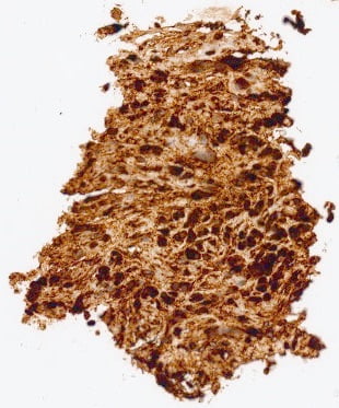
Figure 4. Histological examination: spindle cells arranged in bundles, with dense distribution of nucleus, forming palisades in dense fibrillar stroma; immunohistochemistry staining showing a diffuse cytoplasmic immunoreactivity for S-100 protein.
Schwannomas are autonomic nerve tumors arising from Schwann cells of the neural sheath (1). They comprise 2-8% of all gastrointestinal mesenchymal tumors and are typically located at the stomach. Colonic schwannomas are exceedingly rare. They are mostly diagnosed in the sixth decade, have no gender preponderance and are usually asymptomatic. Symptomatic patients usually complain of constipation, hematochezia or abdominal pain (2). Endoscopically, schwannomas appear as wide-based sessile subepithelial lesions with a rubbery or firm consistency (3). Computed tomography and magnetic resonance imaging do not provide additional information for the distinction from other mesenchymal tumors (1, 3). On EUS, schwannomas typically arise from the second or fourth layer and usually appear as hypoechoic, round or oval, well-demarcated lesions (4), with a similar appearance to leiomyomas and gastrointestinal stromal tumors (GIST). EUS-elastography is a promising tool showing a predominantly hard pattern (5). Taking into account unspecific endoscopic and ultrasonographic features, tissue sampling is required to obtain the definitive diagnosis. However, preoperatively diagnosis is achieved in a minority of cases given the difficulty of obtaining an adequate specimen. When available, EUS-fine needle aspiration or biopsy is the most accurate method for obtaining tissue. Histologically, schwannomas are composed of bundles of spindle cells aligning in a trabecular pattern, surrounded by fibrovascular septa containing lymphoplasmacytic infiltration. On immunohistochemistry, tumor cells are universally positive for S-100 and vimentin but negative for CD117, CD34 and a smooth muscle actin (3). Currently, complete surgical resection is the best therapeutic choice. (1, 6).
REFERENCES
- Barbeiro S, Martins C, Goncalves C, Arroja B, Canhoto M, Silva F, et al. Schwannoma-A Rare Subepithelial Lesion of the Colon. GE Port J Gastroenterol. 2015;22(2):70-4.
- Cakir T, Aslaner A, Yaz M, Gunduz U. Schwannoma of the sigmoid colon. BMJ Case Rep. 2015;2015.
- Chayanupatkul M, Gould Suarez M, Sealock RJ. Colonic Schwannoma Diagnosed by Endoscopic Ultrasound With Fine-Needle Aspiration. Clin Gastroenterol Hepatol. 2018;16(5):A29-A30.
- Faulx AL, Kothari S, Acosta RD, Agrawal D, Bruining DH, Chandrasekhara V, et al. The role of endoscopy in subepithelial lesions of the GI tract. Gastrointest Endosc. 2017;85(6):1117-32.
- Kim S, Yoo I, Kwon C, Hong S, Cho J. Utility of EUS elastography in the diagnosis of gastric subepithelial tumors: a pilot study. Gastrointestinal Endoscopy. 2019.
- Mekras A, Krenn V, Perrakis A, Croner RS, Kalles V, Atamer C, et al. Gastrointestinal schwannomas: a rare but important differential diagnosis of mesenchymal tumors of gastrointestinal tract. BMC Surg. 2018;18(1):47.
AUTHORS
Mafalda João1, Daniel Brito1, Luís Elvas1, José Paulo Magalhães2, Ana Teresa Cadime1.
- Gastroenterology Department, Portuguese Oncology Institute of Coimbra, Portugal
- Pathology Department, Portuguese Oncology Institute of Coimbra, Portugal