
Pesquisa



US Quiz of the Month – junho 2026
Case Report
A 60-year-old female patient presented to the emergency department because of a two-day history of melena associated with progressive asthenia. She denied hematemesis. Laboratory testing revealed iron deficiency anemia (Hb 7.4 g/dL).
Upper endoscopy demonstrated a pedunculated polypoid lesion with congested mucosa and superficial erosions, arising from the duodenal bulb and extending into the third portion of the duodenum. This lesion was suggestive of a subepithelial lesion, presumably responsible for gastrointestinal bleeding (Figure 1).
Endoscopic ultrasound (EUS) identified the lesion on the third layer of the duodenal wall, maintaining a clear interface with the fourth layer with well-defined and regular borders. The peduncle was hypoechoic and had blood vessels inside it, while the head of the lesion was hyperechoic and had blood vessels inside it (Figure 2.a & b & c).
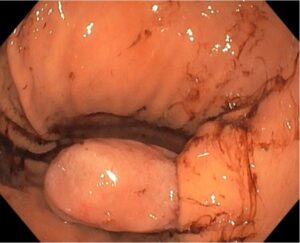
Figure 1 – Upper endoscopy identified a pedunculated polypoid lesion with congested mucosa and superficial erosions, arising from the duodenal bulb and extending into the third portion of the duodenum.
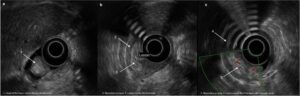
Figure 2 – (a,b,c)- EUS identified the lesion located on the third layer of the duodenal wall, maintaining a clear interface with the fourth layer, with well-defined and regular borders. The peduncle was predominantly hypoechoic and contained internal vascularity, while the head of the lesion was mainly hyperechoic with intralesional vessels.
Based on these endoscopic ultrasound findings, what do you think is the most likely diagnosis?
The lesion was excised by endoloop-assisted snare polypectomy (Figure 3. a & b & c).
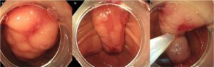
Figure 3 (a,b,c) – The pedunculated lesion, with its base implanted on the duodenal bulb (a/b), was excised using diathermy after securing the pedicle with an endoloop (c).
Histological examination revealed a mature adipocyte proliferation in the submucosa, with focal cellular areas. The cellular areas were composed of bland, short, spindle-shaped cells and ropy collagen bundles admixed with mature adipocytes (Figure 4 a & b). Occasional multinucleated cells and mast cells were observed. Immunohistochemically, the spindle cells stained positive for CD34 (Figure 4. c).
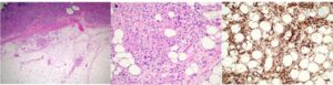
Figure 4 – (a)- Hematoxylin & eosin (HE) 20x. Mature adipocyte proliferation in the submucosa, focally with cellular areas; (b)– HE 200x. Cellular areas are composed of bland short spindled cells and ropy collagen bundles admixed with mature adipocytes. Occasional multinucleated cells and mast cells are seen; (c) Immunohistochemistry (CD34) 200x. The tumor is positive for CD34.
Discussion
Spindle cell lipomas (SCLs) are a morphologically and molecularly distinct subtype of lipoma that most commonly occur in the shoulders, posterior neck, and upper back (1). Their occurrence in the gastrointestinal tract is exceedingly rare, with only a few cases reported in the literature, and involvement of the duodenum is particularly uncommon (2).
SCLs are typically painless, elastic masses ranging from 3 to 5 cm in size, but they can sometimes be larger. Gastrointestinal bleeding caused by a duodenal SCL is extremely rare (3)(4). As illustrated in Figures 1 and 2, gastrointestinal bleeding can be caused by duodenal SCL.
As illustrated in Figure 2 (a/b/c), on endoscopic ultrasonography (EUS), SCLs usually present with smooth margins and well-defined boundaries. They show irregular internal echogenicity with patches and streaks due to a combination of fatty and fibrous components. The presence of internal vascularity can help differentiate them from simple lipomas (5).
Histologically, SCLs are characterized by a combination of mature adipose cells and small, uniform spindle cells set against a background of fibrous stroma (acidophilic collagen fibers) with myxoid changes. Spindle cells typically express CD34 (95%–100%) and are negative for S-100 and smooth muscle actin (SMA)(6), as illustrated in Figure 4 (a/b/c).
SCLs generally have a benign clinical course. In spindle cell lipomas arising in typical anatomical locations, such as the neck and back, the local recurrence rate is approximately 2–3% after complete excision (5). The recurrence rate of duodenal spindle cell lipoma has not been specifically described in the medical literature, likely due to its rarity in this location. There is no standardized or guideline-based follow-up protocol after excision of duodenal SCLs. In asymptomatic patients, follow-up can be individualized, with endoscopic evaluation reserved for those who develop new symptoms.
References
- Enzinger FM, Harvey DA. Spindle cell lipoma. Cancer. 1975 Nov;36(5):1852–9.
- Bancroft LW, Kransdorf MJ, Peterson JJ, Sundaram M, Murphey MD, O’Connor MI. Imaging Characteristics of Spindle Cell Lipoma. American Journal of Roentgenology. 2003 Nov;181(5):1251–4.
- Tao L, Yu H, Zhang L, Liu J, Hu X. A rare case of duodenal spindle cell lipoma with gastrointestinal bleeding. Revista Española de Enfermedades Digestivas. 2022;
- Van Treeck BJ, Fritchie KJ. Updates in spindle cell/pleomorphic lipomas. Semin Diagn Pathol. 2019 Mar;36(2):105–11.
- Fletcher CD, Martin-Bates E. Spindle cell lipoma: a clinicopathological study with some original observations. Histopathology. 1987 Aug;11(8):803-17.
- Templeton SF, Solomon AR. Spindle cell lipoma is strongly CD34 positive. An immunohistochemical study. J Cutan Pathol. 1996 Dec 27;23(6):546–50.
Authors
Sandra Ribeiro Correia1, Ricardo Küttner-Magalhães1,2, Sofia Neves3, Isabel Pedroto1,2, Daniela Gonçalves Ferreira1,2
- Serviço de Gastrenterologia, Unidade Local de Saúde de Santo António
- Instituto de Ciências Biomédicas Abel Salazar, Universidade do Porto
- Serviço de Anatomia Patológica, Unidade Local de Saúde de Santo António