Pesquisa
US Quiz of the Month – maio 2025
Case Report
We present the case of a 71-year-old male patient with past medical history significant for radically resected prostate adenocarcinoma in 2019. In 2023, several bone and lung metastasis developed, for which the patient started hormonotherapy (Darolutamide and Leuprorelin), achieving complete response 8 months later. In 2025, despite PSA blood levels were negative (<0,01ng/dL), a follow-up 68Ga-Prostate-specific membrane antigen (PSMA) PET/TC revealed a de novo nodular uptake in the pancreatic tail (SUV 8,1) (Fig 1.).
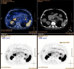
Figure 1. 68Ga-PSMA PET/TC: Nodular uptake in the pancreatic tail.
Abdominal MRI confirmed a solid nodular lesion in the pancreatic tail, with 30x20mm, with a pattern identical to that of the spleen. To exclude an ectopic intrapancreatic accessory spleen, a 99mTc-labelled red blood cell (RCR) scintigraphy was performed, but the pancreatic tail lesion did not show any radiopharmaceutical accumulation suggestive of splenosis (Fig. 2).
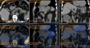
Figure 2. 99mTc-labelled RBR scintigraphy: No radiopharmaceutical accumulation the pancreatic tail.
The patient was then referred to out Digestive Unit to perform an EUS-FNB. On EUS examination, a hypoechoic homogeneous lesion, with well-defined borders, was seen in the pancreatic tail, measuring 17x30mm. A transgastric EUS-FNB was performed using a 22G needle, 2 passes (EchoTip ClearCore EUS Biopsy Needle, Cook Medical) (Video 1).
Video 1. EUS (transgastric view): FNB of pancreatic tail hypoechoic homogeneous lesion, with well-defined borders, measuring 17x30mm.
What is the most likely diagnosis?
Discussion
Pathology showed neoplastic cells positive for CAM 5.2, synaptophysin and chromogranin and negative for KX3.1, consistent with the diagnosis of a well-differentiated neuroendocrine tumor (0 mitosis/mm2, Ki-65 1%) (Fig. 3).
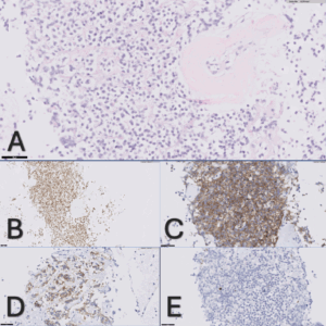
Fig. 3. Pathology: Neoplastic cells (A: H&E, 40X) positive for CAM 5.2 (B, 20X), synaptophysin (C, 40X) and chromogranin (D, 40X), with a Ki-65 of 1% (E, 40x).
A staging PET-DOTANOC was done and showed uptake in the pancreatic tail lesion (SUV max 19,3) and in some peri-centimetric paraaortic lymph nodes (SUV max 6,3) (Fig. 4).
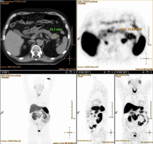
Fig. 4. PET-DOTANOC: Uptake in the pancreatic tail.
During the past years, PSMA PET/CT has become a key imaging modality for the detection, staging and surveillance of prostate adenocarcinoma due to its high sensitivity. PSMA is a transmembrane protein overexpressed not only in prostate epithelial malignant cells but also in endothelial cells of tumor-associated neovasculature of other solid cancers.
To date a few case reports of pNET accidentally diagnosed in PSMA PET/TC have been published. [1-3] Since NETs have very high vascularity, they might show PSMA expression.
Our case report highlights that when PSMA uptake is seen in atypical location a differential diagnosis (other than prostate adenocarcinoma metastasis) needs to be considered, and further evaluation should be performed.
References
- Luong TV, Iversen P, Bouchelouche K, Arveschoug AK. 68Ga-Prostate-Specific Membrane Antigen Uptake in a Pancreatic Neuroendocrine Tumor. Clin Nucl Med. 2020 May;45(5):379-382. doi: 10.1097/RLU.0000000000002997. PMID: 32149792.
- Luna R, Laheru DA, Shin EJ, Lossos C, Robinson MT, Rowe SP, Saad E, Markowski MC. Pancreatic Neuroendocrine Tumor incidentally found on 68Ga-PSMA PET/CT. Nuklearmedizin. 2025 Apr;64(2):177-179. English. doi: 10.1055/a-2511-6595. Epub 2025 Apr 17. PMID: 40245883.
- Chatta R, Tsai HK, Pallod S, Shah H. PSMA PET/CT for Detection of Metastatic Pancreatic Neuroendocrine Tumor. Radiol Imaging Cancer. 2025 Sep;7(5):e250093. doi: 10.1148/rycan.250093. PMID: 40879482.
Authors
Susana Marques1, Miguel Bispo1, Rafaela Rego2, Ricardo Rio-Tinto1, Paulo Fidalgo1, Jacques Devière1,2,3
- Digestive Unit, Champalimaud Foundation, Lisbon, Portugal.
- Pathology Department, Champalimaud Foundation, Lisbon, Portugal.
- Department of Gastroenterology, Hepatopancreatology, and Digestive Oncology, Erasme University Hospital – Université Libre de Bruxelles, Brussels, Belgium.