Pesquisa
US Quiz of the Month – Setembro 2022
CASE REPORT
The authors present the case of a 23-year-old man with recurrent abdominal pain in the right hypochondrium. An abdominal ultrasound detected the presence in the gallbladder of an endoluminal cystic image near the infundibulum, immovable, of ring-like appearance, with a regular wall, measuring about 20 mm (Fig. 1).
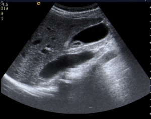
Figure 1 – Abdominal ultrasound: an endoluminal cystic image near the gallbladder’s infundibulum, immovable, of ring-like appearance, with a regular wall, measuring about 20 mm.
An endoscopic ultrasound (EUS) was performed, confirming the presence of a homogeneously echogenic lesion with central low echogenicity, adherent to the gallbladder, measuring 18 mm (Fig. 2).
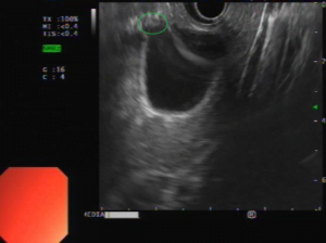
Figure 2 – Endoscopic ultrasound: a homogeneously echogenic lesion with central low echogenicity, adherent to the gallbladder, measuring 18 mm.
The patient was referred for cholecystectomy and a histopathological analysis was performed (Fig. 3).

Figure 3 – Histopathological analysis of the gallbladder.
WHAT IS THE MOST LIKELY DIAGNOSIS?
Pathological analysis revealed a submucosal area of heterotopical gastric mucosa in the gallbladder, comprising mostly of gastric body mucosa and partly of pyloric mucosa. During follow-up for two years the patient was asymptomatic.
Heterotopic gastric mucosa (HGM) is most often localized in the upper intestine but it can be found in the whole gastrointestinal tract [1,2]. Rarely, it can also be localized in the gallbladder [1,2,3]. HGM in the gallbladder is frequently an incidental finding but it can also become symptomatic and can even present as an acute abdomen [1]. Regarding diagnostic imaging, there are no characteristic findings to properly differentiate it from other usual polyps and adenocarcinoma [2]. Although it had been previously considered to have potential for carcinogenesis, there have been no cases reported of malignant transformation [3].
REFERENCES
- Beeskow AB, Meyer H, Schierle K, et al. Heterotopic gastric mucosa in gallbladder – A rare differential diagnosis to gallbladder masses. Medicine. 2018;97:10.
- Hayama S, Suzuki Y, Takahashi M, et al. Heterotopic Gastric Mucosa in the Gallbladder: Report of Two Cases. Surg Today. 2010;40:783–787
- Pendharkar D, Khetrapal S, Jairajpuri ZS, et al. Pancreatic and Gastric Heterotopia in the Gallbladder: A Rare Incidental Finding. Int J Appl Basic Med Res. 2019;9(2):115–117.
AUTHORS
Diogo Bernardo Moura, Nuno Nunes, Francisca Côrte-Real, Carolina Chálim Rebelo, Maria Antónia Duarte.
Gastroenterology Department, Hospital do Divino Espírito Santo de Ponta Delgada, Ponta Delgada, Portugal.